
Treatments · Diagnostic & Therapeutic Injections
Sacroiliac Joint (SI Joint) Injection
An image-guided injection directly into the sacroiliac joint — used both to confirm the joint as the actual source of a patient's pain and, often, to provide direct relief in the same visit.
What it involves
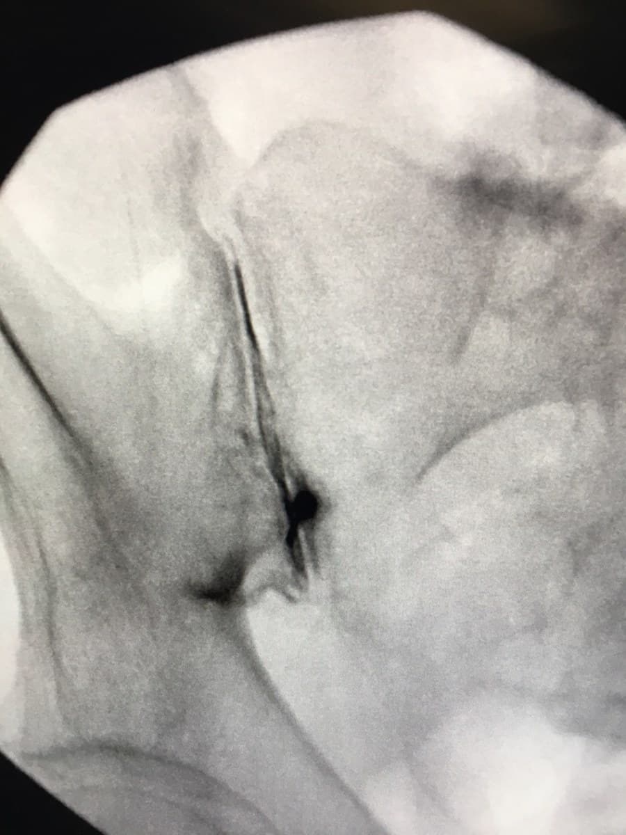
The SI joint sits deep beneath the surrounding muscle and ligament, and its exact location varies enough from patient to patient that reliably placing a needle inside the joint capsule requires image guidance — fluoroscopy or ultrasound — rather than landmarks alone. A local anesthetic, often combined with a corticosteroid, is injected directly into the joint.
Because physical exam maneuvers and imaging alone can't reliably confirm the SI joint as a pain generator, this injection functions as both test and treatment: it directly answers whether the joint is the source, while the anesthetic and steroid combination often provides real, if not always permanent, relief on its own.
How it works
If a patient's pain resolves — or improves substantially — in the distribution typical of SI joint pain while the anesthetic is active, that's strong evidence the joint is the actual pain generator rather than a lumbar disc or facet problem referring pain to the same general area. The corticosteroid component then reduces inflammation within the joint capsule, which can extend relief well beyond the anesthetic's effect.
For patients who get clear but temporary relief, that result directly shapes what comes next — supporting a case for radiofrequency ablation of the nerves supplying the joint, or, for a confirmed, structurally dysfunctional joint that hasn't held up to injections, SI joint fusion.
How We Build Your Plan
The clinical reasoning behind this recommendation
What's the diagnosis?
A history and physical exam pattern suggestive of SI joint involvement — pain typically felt just below the belt line on one side — confirmed directly by an image-guided injection rather than exam maneuvers or imaging alone.
What's the mechanism?
Local anesthetic delivered precisely into the SI joint capsule temporarily interrupts its pain signaling; corticosteroid added to the same injection reduces inflammation within the joint, extending relief beyond the anesthetic's effect alone.
What are the options?
The injection can be diagnostic alone, therapeutic on its own for some patients, or the first step before a more durable option like radiofrequency ablation or SI joint fusion, depending on how the patient responds and how long relief lasts.
Why this approach?
Because the SI joint is difficult to isolate on exam and rarely shows definitively on standard imaging, Dr. Foster uses this injection as the deciding step — confirming the joint as the pain generator before recommending anything more invasive.
What are the tradeoffs?
The injection is low-risk and provides valuable diagnostic information regardless of outcome, but relief — even with the added steroid — isn't always durable. A clear, reproducible response is what supports moving toward a longer-lasting option.
What to Expect
A brief, image-guided outpatient procedure
The injection itself typically takes about 5 minutes, done under fluoroscopic or ultrasound guidance on an outpatient basis. Patients track how much their usual pain improves over the next several hours while the anesthetic is active — the window that confirms the joint as the source.
Mild soreness at the injection site is common for a day or two. When effective, relief can last anywhere from several weeks to several months; if it fades sooner than hoped, the result still guides the next step in the care pathway.
Conditions This Treats
Conditions this treatment addresses
Think your pain might be coming from the SI joint?
An accurate diagnosis starts with a conversation. Reach out to schedule a consultation.